BPArt: Showcasing the Microscopic Beauty of Brain Research
To help populate our new website, we invited members of the Biological Psychiatry Australia community to submit original images that highlight the beauty and complexity of their research.
This one-time competition celebrated every submission as a winner—each image offering a unique glimpse into the cellular and structural intricacies of the brain. From fluorescent neurons to intricate tissue landscapes, these works showcase the visual power of science at a microscopic scale.
We’re proud to display this collection as a testament to the creativity and curiosity that drive our field, and thank the researchers that submitted their work.
Cassandra Hoffmann
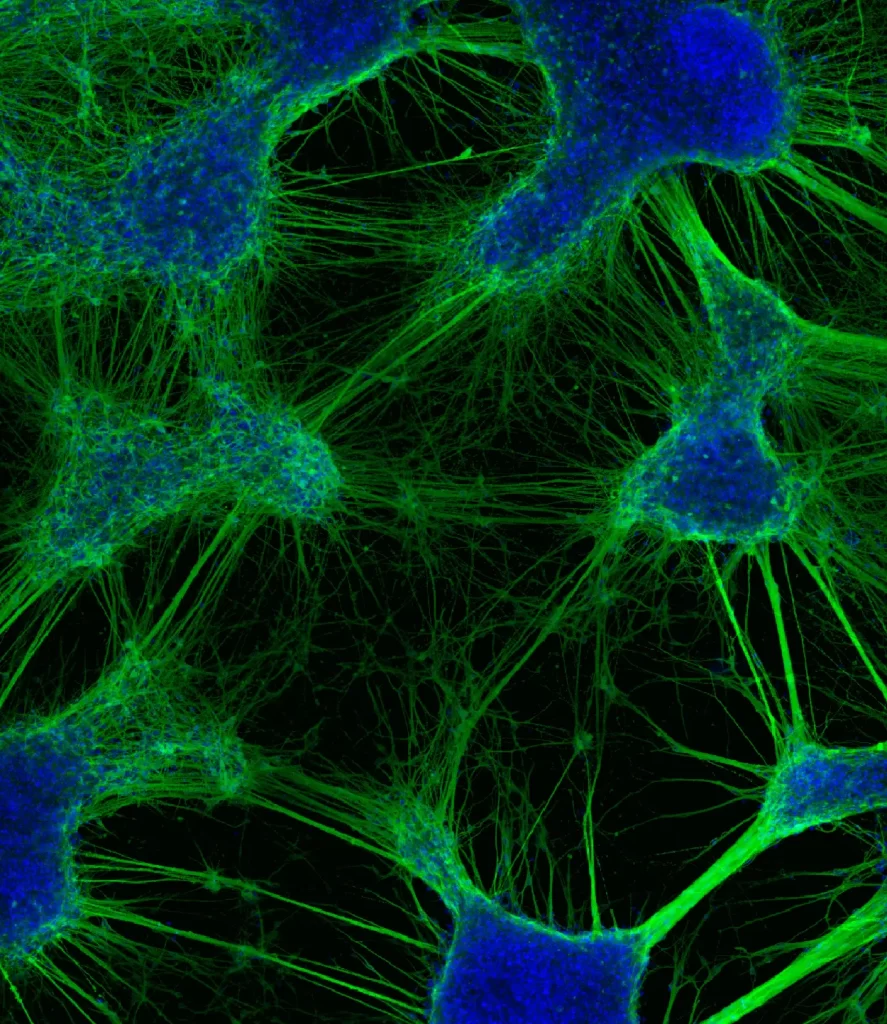
Human stem cell-derived neurons organise into efficient networks in vitro.
Image stack captured with LSM900 confocal at 10x. Neuron cytoskeleton stained with anti-beta tubulin and Alexa Fluor 488 (green) and nuclei counterstained with DAPI (blue).
Cassandra Hoffmann1*, Ellie Cho2, Andrew Zalesky1,3, Maria A. Di Biase1,4,5
1.Systems Neuroscience Lab, Melbourne Neuropsychiatry Centre, Department of Psychiatry, The University of Melbourne, Parkville, Australia
2.Biological Optical Microscopy Platform, University of Melbourne, Parkville, Australia
3.Department of Biomedical Engineering, The University of Melbourne, Parkville, Australia
4.Stem Cell Disease Modelling Lab, Department of Anatomy and Physiology, The University of Melbourne, Parkville, Australia
5.Psychiatry Neuroimaging Laboratory, Department of Psychiatry, Brigham and Women’s Hospital, Harvard Medical School, Boston, United States
Publication link: https://www.nature.com/articles/s42003-024-06264-9
Jordan Clarke
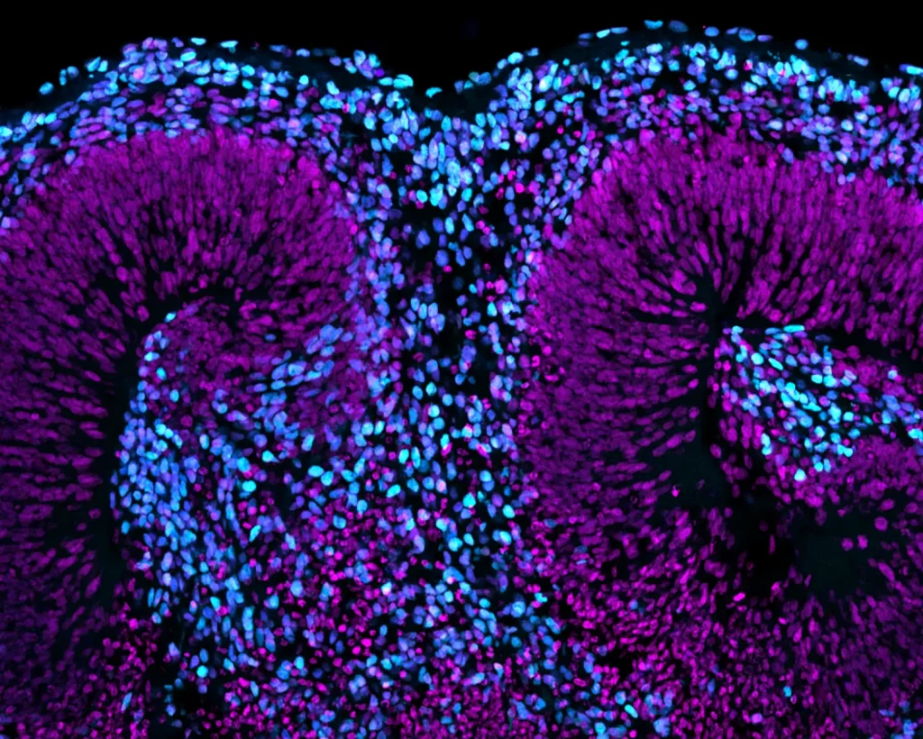
Day 40 Cortical Organoid section showing neural rosette formation (nuclei, MAGENTA) and expression of TBR1 (CYAN) and CTIP2 (BLUE) indicitive of early deep-layer neuron and immature preplate development. Taken on the LSM900 ZEISS confocal at 10X.
Jordan E. Clarke 1, Angela Connelly 2, Maciej Daniszewski 1, Yumiko Hirokawa 1, Alice Pébay 1,3, Murray Cairns 4,5 & Maria A. Di Biase 1,6,7
1 Stem Cell Disease Modelling Lab, Department of Anatomy and Physiology, The University of Melbourne, Australia
2 Central Cardiovascular Regulation Laboratory, Department of Anatomy and Physiology, The University of Melbourne, Australia
3 Department of Surgery, Royal Melbourne Hospital, Melbourne Medical School, The University of Melbourne, Melbourne, VIC, Australia
4 School of Biomedical Sciences and Pharmacy, University of Newcastle, Newcastle, NSW, Australia
5 Centre for Brain and Mental Health Research, Hunter Medical Research Institute, Newcastle, NSW, Australia
6 Systems Neuroscience Lab, Melbourne Neuropsychiatry Centre, Department of Psychiatry, The University of Melbourne, Australia
7 Psychiatry Neuroimaging Laboratory, Department of Psychiatry, Brigham and Women’s Hospital, Harvard Medical School, United States
STEM CELL DISEASE MODELLING LAB https://biomedicalsciences.unimelb.edu.au/sbs-research-groups/anatomy-and-physiology-research/stem-cell-and-developmental-biology/pebay-lab-stem-cell-disease-modelling#details
Bruna Panizzutti Parry
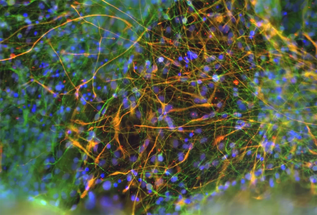
Illuminating Bipolar Disorder: A Neural Symphony
A vibrant co-culture of iPSC-derived neurons (green, MAP2) and astrocytes (orange, GFAP) from participants with bipolar disorder. This dynamic network reveals the intricate cellular interplay within the brain, offering a window into the neurobiological foundations of mood disorders.
Bruna Panizzutti, Megan Ellis, Courtney Swinton, Ken Walder
Berk, M., Kim, J. H., Williams, L. J., Liu, Z. S. J., Siskind, D., Panizzutti, B., Yung, A. R., & Walder, K. A novel discovery platform for targeted drug repurposing: application for psychiatric disorders. The Lancet Psychiatry. https://doi.org/10.1016/S2215-0366(25)00066-5
Deakin University, School of Medicine, IMPACT, The Institute for Mental and Physical https://impact.deakin.edu.au/
Brandon Richards
This fluorescent photomicrograph depicts a coronal mouse brain section immunostained for the dopamine marker tyrosine hydroxylase (pink) and calcium-binding protein parvalbumin (cyan). This image particularly highlights a cluster of tyrosine hydroxylase-expressing cells in the hypothalamus known as the A13 dopamine group, observed bilaterally in the middle of the image. These cells are of particular interest to our group given their role in aberrant fear expression – a hallmark of pervasive fear-related psychopathologies such as post-traumatic stress disorder.
Brandon Richards – Macquarie University
Christina Perry – Macquarie University
Jordan Clarke
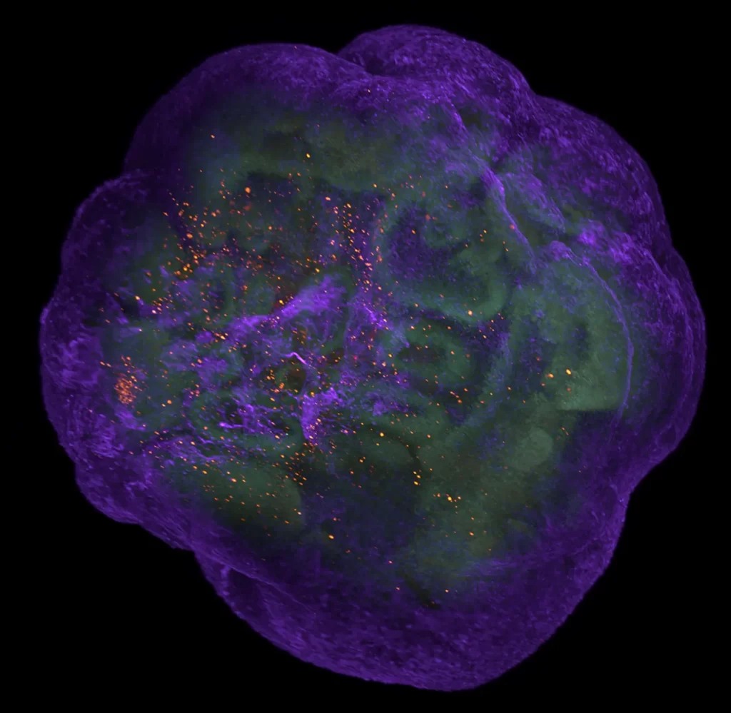
D40 wholemount Cortical Organoid showing spatial organisation of neural rosettes (nuclei: PINK, intermediate & glial progenitors: ORANGE), early neuron formation (neural cytoskeletons: PURPLE), early microglia/microglial progenitors (CYAN) and a centralised core of radial glia (GREEN). Taken on Miltenyi UltraMicroscope Blaze Light Sheet at 12X, using fast tiles. Organoid is approx. 2.5mm in size.
Jordan E. Clarke 1, Alice Pébay 1,2, Murray Cairns 3,4 & Maria A. Di Biase 1,5,6
1 Stem Cell Disease Modelling Lab, Department of Anatomy and Physiology, The University of Melbourne, Australia
2 Department of Surgery, Royal Melbourne Hospital, Melbourne Medical School, The University of Melbourne, Melbourne, VIC, Australia
3 School of Biomedical Sciences and Pharmacy, University of Newcastle, Newcastle, NSW, Australia
4 Centre for Brain and Mental Health Research, Hunter Medical Research Institute, Newcastle, NSW, Australia
5 Systems Neuroscience Lab, Melbourne Neuropsychiatry Centre, Department of Psychiatry, The University of Melbourne, Australia
6 Psychiatry Neuroimaging Laboratory, Department of Psychiatry, Brigham and Women’s Hospital, Harvard Medical School, United States
STEM CELL DISEASE MODELLING LAB
.
Jordan Clarke
D40 wholemount Cortical Organoid showing multiple neural rosette formations (neural stem cells/early neural & glial progenitors (GREEN), early immature neuron development and organisation (PURPLE) and a population of integrated microglia progenitors (ORANGE). Taken on Miltenyi UltraMicroscope Blaze Light Sheet at 4X. Organoid is approx. 2.5mm in size.
Jordan E. Clarke 1, Alice Pébay 1,2, Murray Cairns 3,4 & Maria A. Di Biase 1,5,6
1 Stem Cell Disease Modelling Lab, Department of Anatomy and Physiology, The University of Melbourne, Australia
2 Department of Surgery, Royal Melbourne Hospital, Melbourne Medical School, The University of Melbourne, Melbourne, VIC, Australia
3 School of Biomedical Sciences and Pharmacy, University of Newcastle, Newcastle, NSW, Australia
4 Centre for Brain and Mental Health Research, Hunter Medical Research Institute, Newcastle, NSW, Australia
5 Systems Neuroscience Lab, Melbourne Neuropsychiatry Centre, Department of Psychiatry, The University of Melbourne, Australia
6 Psychiatry Neuroimaging Laboratory, Department of Psychiatry, Brigham and Women’s Hospital, Harvard Medical School, United States
STEM CELL DISEASE MODELLING LAB:
https://biomedicalsciences.unimelb.edu.au/sbs-research-groups/anatomy-and-physiology-research/stem-cell-and-developmental-biology/pebay-lab-stem-cell-disease-modelling#details
Jordan Clarke
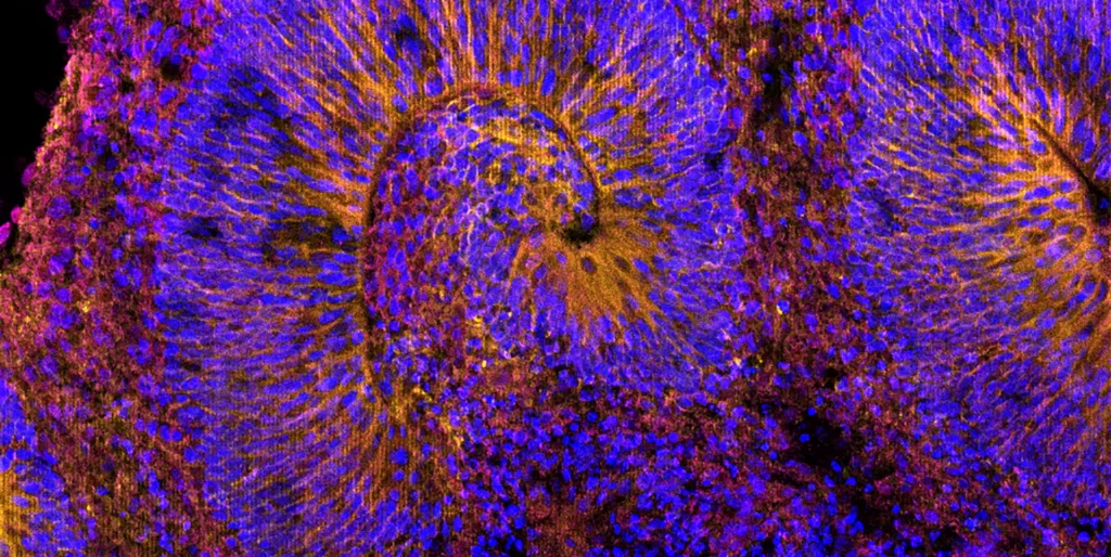
Day 40 Cortical Organoid section displaying neural rosette formation (nuclei, BLUE) and development of radial glia and early-intermediate neural progenitors (GLAST-1: YELLOW, TUJ1: MAGENTA). Taken on the LSM900 ZEISS confocal at 10X.
Jordan E. Clarke 1, Angela Connelly 2, Maciej Daniszewski 1, Yumiko Hirokawa 1, Alice Pébay 1,3, Murray Cairns 4,5 & Maria A. Di Biase 1,6,7
1 Stem Cell Disease Modelling Lab, Department of Anatomy and Physiology, The University of Melbourne, Australia
2 Central Cardiovascular Regulation Laboratory, Department of Anatomy and Physiology, The University of Melbourne, Australia
3 Department of Surgery, Royal Melbourne Hospital, Melbourne Medical School, The University of Melbourne, Melbourne, VIC, Australia
4 School of Biomedical Sciences and Pharmacy, University of Newcastle, Newcastle, NSW, Australia
5 Centre for Brain and Mental Health Research, Hunter Medical Research Institute, Newcastle, NSW, Australia
6 Systems Neuroscience Lab, Melbourne Neuropsychiatry Centre, Department of Psychiatry, The University of Melbourne, Australia
7 Psychiatry Neuroimaging Laboratory, Department of Psychiatry, Brigham and Women’s Hospital, Harvard Medical School, United States
STEM CELL DISEASE MODELLING LAB
Jordan Clarke
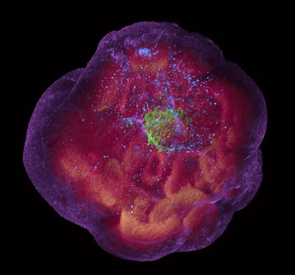
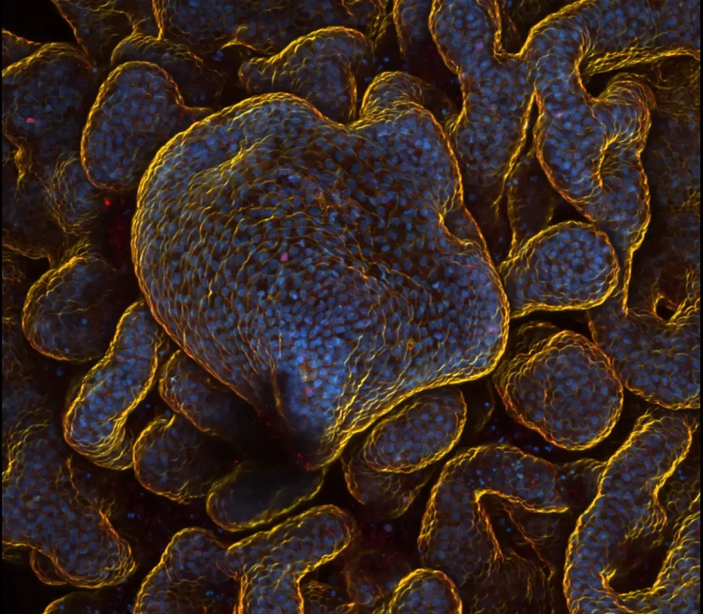
Close-up, z-projection of a Choroid Plexus organoid exhibiting the typical papillary protuberances of the specialised ChP epithelium (nuclei – HOECHST: BLUE, Phalloidin: YELLOW, PAX6: RED). Taken on the LSM900 ZEISS confocal at 10X, 135 steps, 2.5uM step size. (This organoid developed after an embryoid body failed to undergo proper neural induction and form cortical tissue).
Jordan E. Clarke 1, Alice Pébay 1,2, Murray Cairns 3,4 & Maria A. Di Biase 1,5,6
1 Stem Cell Disease Modelling Lab, Department of Anatomy and Physiology, The University of Melbourne, Australia
2 Department of Surgery, Royal Melbourne Hospital, Melbourne Medical School, The University of Melbourne, Melbourne, VIC, Australia
3 School of Biomedical Sciences and Pharmacy, University of Newcastle, Newcastle, NSW, Australia
4 Centre for Brain and Mental Health Research, Hunter Medical Research Institute, Newcastle, NSW, Australia
5 Systems Neuroscience Lab, Melbourne Neuropsychiatry Centre, Department of Psychiatry, The University of Melbourne, Australia
6 Psychiatry Neuroimaging Laboratory, Department of Psychiatry, Brigham and Women’s Hospital, Harvard Medical School, United States
STEM CELL DISEASE MODELLING LAB